Course Case Studies
- Back to Course Home
- Participation Instructions
- Review the course material online or in print.
- Complete the course evaluation.
- Review your Transcript to view and print your Certificate of Completion. Your date of completion will be the date (Pacific Time) the course was electronically submitted for credit, with no exceptions. Partial credit is not available.
Patient BB is a woman, 74 years of age, who presents to her physician's office with complaints of dizziness and recent unexpected falls. She reports that she experiences symptoms once every week or so. When questioned about her falls, she states that she thinks she "blacks out for a second" but "comes to fast" and "knows where she is." She denies any injury from the falls. On admission, her blood pressure is 154/86 mm Hg, her heart rate is 68 bpm and irregular, and her respiratory rate is 14. Patient BB is alert and oriented, and does not appear to be in any distress. A 12-lead ECG shows that the patient is in atrial fibrillation at a controlled rate. A review of Patient BB's medical record shows that she has persistent atrial fibrillation. Past attempts at pharmacologic and electrical cardioversion have failed, and Patient BB remains on low dose antiarrhythmic therapy for rate control and on warfarin for anticoagulation. She has a history of hypertension, coronary artery disease, and percutaneous transluminal angioplasty (PTCA) for placement of two stents. She also has a history of a mitral valve replacement with an artificial valve. Patient BB lives alone in a senior citizen apartment complex; she participates in the social life of the community and takes frequent walks outside in good weather. Her favorite hobby is window-shopping at the mall.
Comments and Rationale: Complaints of dizziness and frequent, unexplained falls are symptoms commonly associated with symptomatic bradycardia. Symptomatic bradycardic episodes are often transient and difficult to capture either on 12-lead ECG or during routine vital sign checks. Due to Patient BB's age, she is at risk to develop symptomatic bradycardia as a result of age-related changes in the cardiac conduction system. She is also more likely to develop conduction abnormalities from antiarrhythmic therapy and medications prescribed to manage hypertension.
The physician orders AECG monitoring for Patient BB. A continuous loop event recorder is selected, and the patient is instructed on its use. She is also instructed to contact the physician when she has experienced one of her symptomatic episodes.
Comments and Rationale: A continuous loop event recorder may be used to document arrhythmias that occur relatively infrequently. This type of AECG is worn continuously; the patient is instructed to depress a switch when he or she feels symptoms. Although Patient BB appears to lose consciousness briefly during her episodes, she is not severely disoriented when she returns to consciousness and would be able to depress the switch in time to signal the device to save the data. Data from the AECG may be transmitted via telephone for analysis.
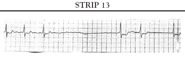
Approximately 10 days after Patient BB begins wearing the AECG, she experiences another symptomatic episode. Following instructions, she depresses the switch on the recorder. When the episode has ended, she contacts her physician and follows instructions to transmit the data from the AECG via telephone. Analysis of the transmitted data shows that Patient BB is experiencing episodes of severe bradycardia; her ECG tracing shows that she has pauses that last several seconds without an effective heartbeat; during these episodes, her heart rate drops to well less than 60 bpm. See Strip 13.
A diagnosis of symptomatic bradycardia caused by atrial fibrillation with a slow ventricular response is established. Patient BB is referred for pacemaker implantation. Based on a thorough assessment, the physician chooses to implant a VVIR pacemaker.
Comments and Rationale: The definitive diagnosis of symptomatic bradycardia requires that the patient's symptoms of decreased cerebral blood flow and bradyarrhythmia be correlated and documented. For some persons with atrial fibrillation and a slow ventricular response, modifying the prescribed antiarrhythmic therapy may be sufficient to resolve the bradycardic episodes. However, this patient's antiarrhythmic therapy is already low dose and cannot be effectively reduced or altered. Implantation of a permanent pacemaker is indicated. A VVI (or VVIR) is the pacemaker of choice for persons with chronic atrial fibrillation; because a VVI pacemaker does not sense or pace in the atria, it cannot be confused by the chaotic atrial activity that occurs in atrial fibrillation. As Patient BB has an active lifestyle, the rate adaptive feature could be indicated. This will allow the pacemaker to pace her heart at a higher rate when she is engaged in increased activity.
The physician instructs Patient BB to discontinue taking her warfarin two days in advance of her admission to the hospital. When the patient is admitted to the hospital, her INR remains elevated. A continuous heparin drip is initiated and partial thromboplastin times (PTTs) are monitored; the heparin drip is adjusted to maintain the PTT in the desired range. Preoperative lab work, ECG, and chest x-ray are completed. Preoperative education is begun. Two days after admission, Patient BB's INR has dropped to the desired level, and pacemaker implantation is scheduled for the next day. Preoperative preparation is completed; Patient BB is made NPO overnight, and her heparin drip is discontinued. The following morning, Patient BB is taken to the cardiac catheterization lab. Under sedation and local anesthesia, the pacemaker is implanted. The low rate setting is programmed at 60 bpm, and the upper rate limit for rate adaptation is set at 120 bpm.
Comments and Rationale: To reduce the risk of bleeding during and after implantation, warfarin is discontinued prior to the procedure, and the patient's INR is allowed to return to near normal. However, because of Patient BB's chronic atrial fibrillation and artificial heart valve, she is at high risk for thromboembolic events. To reduce this risk, she is admitted to the hospital several days before the implantation is scheduled and placed on a heparin drip. To limit the risk of bleeding during the procedure, the heparin drip is discontinued four to six hours before the procedure. However, as previously stated, a strategy of implanting devices during uninterrupted warfarin therapy appears to have a lower bleeding risk than a strategy of temporarily discontinuing warfarin and bridging with heparin. Because the patient has no special needs, the cardiac catheterization lab is an appropriate site for the pacemaker implantation.
Following Patient BB's return to her room, she is placed on continuous telemetry monitoring. Her vital signs are monitored periodically, and the pacemaker pocket site is inspected for signs of excessive swelling or bleeding. She is instructed to limit use of the arm on the same side as the pacemaker generator, and she receives analgesic medications as prescribed for pain management. A chest x-ray is done, and a 12-lead ECG (with and without magnet) is completed. Patient BB's vital signs remain stable; the pacemaker generator site shows no signs of excessive bleeding or swelling. Telemetry monitoring obtains Strip 14.
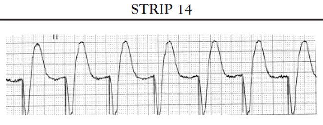
Comments and Rationale: The most common complications in the immediate postoperative period are bleeding or swelling in the pacemaker pocket and accidental dislodgment of the pacemaker lead. A chest x-ray is performed to verify lead position; a 12-lead ECG is used to evaluate pacemaker function. Strip 14 shows normal VVIR pacemaker function. The strip shows a heart rate of approximately 70 bpm; this falls within the lower and upper rate limits of Patient BB's pacemaker. Ventricular pacer spikes are present in front of each QRS complex. The QRS complex shows the typical configuration for a paced QRS. Each pacer spike is followed by a paced QRS; there are no random pacemaker spikes or excessive pauses between beats.
Prior to Patient BB's discharge, she receives information on incision line care and precautions to consider. She receives her temporary pacemaker identification card and is instructed to carry the identification card at all times. She is taught how and when to count her pulse and when to notify the physician. The nurse provides the patient with additional written materials, including the patient education handbook supplied by the pacemaker manufacturer. Patient BB expresses concerns about using cellular phones and flying; the nurse provides specific information about the use of cellular phones and how to manage airport security. Patient BB is discharged home; she has a follow-up appointment one week following discharge.
Comments and Rationale: The major focus of patient education just prior to discharge is on prevention of infection and prevention of accidental dislodgment of the pacemaker leads. Specific patient and family concerns should be addressed. Because patients and families frequently think of additional questions following the patient's discharge, the use of written materials provides a resource for them to use at home. Additional patient education will occur during the follow-up visit.
Patient CC, a man 56 years of age who has a DDD pacemaker, calls the TTM center to report that his heart rate is 30 bpm and that his preimplantation symptoms of dizziness, shortness of breath, and light-headedness have reoccurred. Patient CC's pacemaker was implanted approximately two years earlier for management of third-degree heart block. The rate settings on the DDD pacemaker are 60 bpm (low) and 120 bpm (high). Up to this point, the patient has experienced no problems with his pacemaker. Following instructions from the TTM center, Patient CC transmits a recording of his heart rhythm without application of a magnet.
Comments and Rationale: A heart rate that drops to dramatically less than the low rate setting coupled with a return of preimplantation symptoms is indicative of pacemaker malfunction. Malfunctions that may occur during the mid-life of a pacemaker include damage to the pacemaker lead or its insulation. TTM may be used to identify indications of pacemaker malfunction.
The TTM center receives Strip 15. The center immediately notifies Patient CC to report to the emergency department of his local hospital.
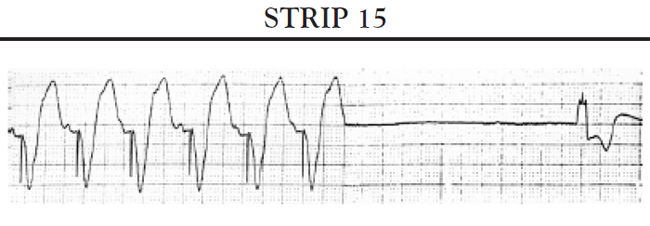
Comments and Rationale: Strip 15 shows a pause of approximately three seconds. The pause is followed by an escape beat. As there are no pacemaker spikes present during the pause, the pacemaker malfunction appears to be failure to fire. Patient CC should obtain immediate medical care.
Upon Patient CC's arrival in the emergency department, he is placed on continuous telemetry monitoring. The monitoring initially shows a heart rate of 60 bpm. Telemetry interrogation of the pacemaker shows indications of probable lead fracture. A chest x-ray confirms the presence of a lead failure. The patient is admitted to a telemetry unit for monitoring and revision of his pacemaker. On the acute care unit, Patient CC is again placed on continuous telemetry monitoring. Telemetry monitoring shows a paced rhythm with intermittent missed beats and an isolated two- to three-second pause. An external noninvasive pacemaker is made available. Preoperative lab work is completed, and the patient is kept NPO. Patient CC is taken to the catheterization lab, and the broken lead is replaced.
Comments and Rationale: Telemetry interrogation can retrieve data that reflects the functioning of the pacemaker leads. Fracture of a pacemaker lead may often be seen on a PA and lateral chest x-ray. Fracture of a lead may result in the pacemaker's inability to deliver a pacing impulse to the heart when needed. Due to the length of Patient CC's pauses and the bradycardic rate of his escape rhythm, equipment for externally pacing the heart should be readily available. Replacement of the pacemaker lead is indicated to correct the pacemaker malfunction.
Following revision of the lead, the patient returns to the inpatient unit. His vital signs are monitored periodically, and the generator pocket site observed for bleeding or swelling. He is placed on continuous telemetry monitoring, and his rhythm is monitored for signs of proper pacemaker functioning. His rate settings remain at 60 bpm (low) and 120 bpm (high). Strip 16 is obtained and documented.
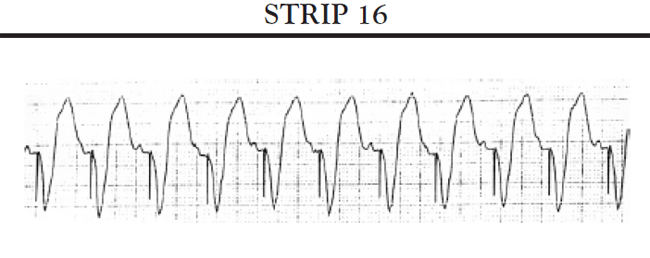
Comments and Rationale: Strip 16 shows normal DDD pacemaker functioning. The patient's SA node is firing normally, and the pacemaker is pacing the ventricles at the same rate. The heart rate is approximately 100 bpm, which is somewhat high but within the rate limit setting of the pacemaker. Each pacemaker spike falls at the appropriate place, and each spike is followed by a QRS complex. Each QRS complex shows the typical configuration for a paced QRS complex. There are no random pacer spikes, and there are no pauses that exceed the interval allowed by the timing interval for the low rate limit setting.
As previously discussed, Patient N, a man 66 years of age, presented to his physician's office complaining of increased shortness of breath, rapid weight gain in the last three to four days, and an inability to perform his usual daily activities due to fatigue. Patient N has a history of MIs, coronary artery bypass surgery, acute coronary syndrome, multiple stent placements, and heart failure. Until recently, Patient N's heart failure has been well managed through the use of medications and diet. However, in the last month, the patient has been admitted to the local hospital twice for management of his heart failure symptoms. Based on an assessment of Patient N's status, the physician decides to admit him to the hospital for further management of his heart failure and evaluation for possible biventricular pacemaker implantation.
Comments and Rationale: Heart failure is a progressive, debilitating disease characterized by increasing symptoms, loss of function, and frequent admissions to the hospital for symptom management. Research has shown that neurohormonal mechanisms in the body contribute to this pattern of increased symptoms and worsening disease. Biventricular pacing has been found to be an effective therapy for the management of heart failure in selected patients.
Patient N's admitting ECG reveals that he is in normal sinus rhythm, with a QRS duration of 150 ms. An echocardiogram shows an ejection fraction of 25% as well as abnormal findings consistent with ventricular dyssynchrony. Based on the patient's symptoms, the physician determines that he is in NYHA functional class III heart failure. A review of Patient N's medications shows that he is receiving optimal doses of recommended medications for the management of heart failure. The physician recommends the implantation of a biventricular pacemaker ICD.
Comments and Rationale: Patient N meets the eligibility criteria for implantation of a biventricular pacemaker: an ejection fraction of 35% or less, NYHA Class III to Class IV heart failure, symptomatic despite optimal medical therapy, and a QRS duration of greater than 120 ms. In addition, the patient's ECG demonstrates additional clinical findings that are indicative of ventricular dyssynchrony. Patient N also meets criteria, based on clinical trial data, for the implantation of an ICD: has a history of ischemic heart disease, is at least 40 days post MI, has a left ventricle ejection fraction less than or equal to 30%, NYHA class II or III symptoms despite optimal medical therapy, and can reasonably be expected to survive with a good functional status for more than one year.
Patient N undergoes successful transvenous implantation of a biventricular pacemaker ICD in the cardiac catheterization lab. Upon return to the floor, his vital signs are stable. He is drowsy but responds appropriately to verbal stimuli. The pacemaker pocket shows no signs of bleeding or hematoma formation. Continuous telemetry monitoring is initiated and Strip 17 is obtained.
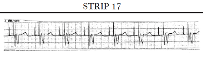
Comments and Rationale: Strip 17 does not show any evidence of pacing problems; each beat is initiated by a P wave, and each beat is followed by a paced QRS complex. Every QRS is paced, and the rate falls within the rate setting programmed for the pacemaker.
Key points of care following implantation of a biventricular pacemaker ICD include assessment of vital signs, continuous monitoring of ECG rhythm, and assessment of the pacemaker pocket for signs of bleeding or hematoma formation. Although definitive diagnosis of pacing problems requires the expertise of a specially trained physician, continuous telemetry monitoring can identify clues that suggest a problem might exist.
About four hours after Patient N's return from the catheterization lab, he indicates that he has become extremely short of breath. His status is assessed. His blood pressure remains within his usual range. His heart rate is 100 bpm, and he remains 100% ventricularly paced on telemetry. His oxygen saturation has fallen to 88% on 2 liters nasal cannula. He has increased crackles and wheezes bilaterally in his lung bases. The physician is notified and orders a portable chest x-ray and a dose of IV furosemide. Patient N verbalizes distress regarding the recurrence of his symptoms, asking if this means "the pacemaker is not going to help."
Comments and Rationale: Shortness of breath following pacemaker implantation in a patient with severe heart failure may be an indication that a pneumothorax has developed as a complication of the procedure, or it may indicate that the patient is experiencing some volume/fluid overload. A chest x-ray can be used to confirm or rule out the presence of a pneumothorax. IV furosemide is a treatment of choice of an acute episode of volume overload. Volume overload postimplantation in a patient with heart failure may occur from a combination of factors, including physiologic stress of the procedure, anxiety, disruption in normal medication times, and the administration of even small amounts of IV fluid during the procedure. It is appropriate to reassure the patient that the occurrence of his symptoms at this point in his hospitalization is not an indication that CRT will be ineffective.
In response to the IV furosemide, Patient N diureses well and his acute episode of dyspnea resolves. He develops no further acute episodes and is discharged home two days later. In addition to the usual discharge instructions for patients with newly implanted pacemakers, Patient N is instructed to adhere to his prescribed medication therapy, diet restrictions, and lifestyle changes. He is advised to identify the level of his symptoms and activity; it is suggested that he keep a diary noting even small improvements. He is instructed to report increasing signs and symptoms of heart failure to his physician and to make sure to keep his follow-up appointments for the evaluation of his device. Because Patient N also has an ICD, specific ICD discharge instructions are also included.
Comments and Rationale: Persons with biventricular pacemakers should understand that the pacemaker does not replace previously prescribed medical therapies, and that just because their symptoms decrease, they cannot stop taking their medications or adhering to their diet restrictions and recommended lifestyle changes. Improvement in activity tolerance and symptom reduction may occur very gradually. Instructing the patient to note his/her baseline level of symptoms/activity and to record every improvement, no matter how small, can encourage the patient that CRT is working. Recurring or increased signs of failure can be an indication that resynchronization has been lost and should be reported immediately to the physician.
At Patient N's six-month follow-up appointment, he tells the physician that he has noticed little improvement in the amount of activity that he can do. With questioning, the patient admits that he has not been re-hospitalized for management of his heart failure symptoms in the last six months and that his ICD has not fired. The physician interrogates the device and obtains an echocardiogram to evaluate the effectiveness of the programming. The physician also reviews Patient N's current medications, with special emphasis on the dosages of his beta blockers and ACE inhibitors. The physician also questions the patient about his daily activities and what steps he has taken to gradually improve his physical conditioning. Based on this assessment data, the physician decides to increase Patient N's ACE inhibitor dose and to prescribe a structured activity program designed to increase his exercise tolerance.
Comments and Rationale: Some people respond more slowly than others to biventricular pacing. Measures that can improve the patient's response include "fine tuning" the device's programming, decreasing symptoms by increasing neurohormonal blockade through increased medication doses, and employing measures to help the patient slowly, carefully increase his/her activity in small steps.
- Back to Course Home
- Participation Instructions
- Review the course material online or in print.
- Complete the course evaluation.
- Review your Transcript to view and print your Certificate of Completion. Your date of completion will be the date (Pacific Time) the course was electronically submitted for credit, with no exceptions. Partial credit is not available.